Knee Injuries
Anterior Cruciate Ligament Tear
LESÕES
DO JOELHO
INFEÇÕES OSTEOARTICULARES

Knee Injuries - Knee Traumatology

Anterior Cruciate Ligament Tear
What is the Anterior Cruciate Ligament?
The Anterior Cruciate Ligament (ACL) is one of the key ligaments in the knee that is crucial for maintaining knee stability. A ligament is a structure that connects two bones, restricting joint mobility. The primary function of the ACL is to prevent excessive forward movement of the tibia.
The ACL also aids in stabilizing the knee in other movements, such as angulation and rotation. Other principal ligaments in the knee include the Lateral and Medial Collateral Ligaments, and the Posterior Cruciate Ligament.
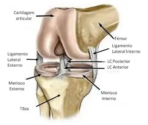
Caption 3: Normal knee ligament anatomy
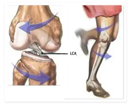
Caption 4: Diagram of the typical injury mechanism
Causes
An ACL tear can occur in anyone but is undoubtedly more common among athletes (e.g., football players). Approximately 70% of these tears result from non-contact injuries, typically through a mechanism where the knee twists, causing the femur and tibia to rotate in opposite directions.
Symptoms
The most frequent symptom is instability or the sensation that the knee is going to collapse. This feeling can vary in intensity and can hinder actions that require knee rotation, sudden changes of direction, or rapid decelerations.
This impairment is particularly noticeable in individuals engaged in sports or physically demanding occupations.
Other symptoms may include recurrent episodes of swelling and pain, or even transient joint locking, often linked to concurrent injuries (e.g., to the menisci or cartilage).
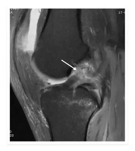
Caption 5: Appearance of an ACL tear on magnetic resonance imaging
Diagnosis
Diagnosis is based on clinical history, physical examination (specific manoeuvres that demonstrate the presence of anteroposterior instability), and findings from diagnostic tests.
The optimal examination to confirm the presence of an ACL tear is magnetic resonance imaging (MRI), which also provides detailed information about the condition of other ligaments, menisci, and articular cartilage.
Treatment Options
Surgical treatment is not always necessary, particularly in sedentary patients and those without associated injuries.
When opting for surgical treatment, the objective is to reconstruct the ligament, also known as ligamentoplasty. There are numerous graft options available. The most common are the patellar tendon (or bone-tendon-bone) and the hamstring tendons, each with its own advantages and disadvantages, though none has demonstrated a clear superiority over the others.
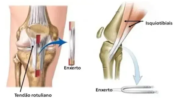
Caption 6: Diagram of patellar graft on the left and hamstring tendons on the right
During surgery, bone tunnels are created in the femur and tibia (replicating the original insertions of the ACL) through which the graft is threaded and then secured.
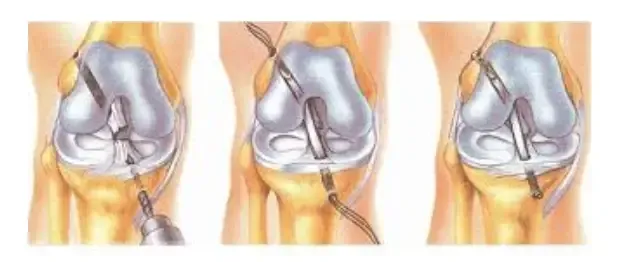
Esquema de colocação de enxerto nos túneis na tíbia e fémur
Currently, this surgery is typically performed via arthroscopy, which entails less surgical trauma and is associated with excellent outcomes in terms of symptomatic relief.

Fotografia inter-operatória após reconstrução artroscópica do LCA


Knee
Osteoarthritis
Is a chronic and progressive disease that can affect any joint in the body
Click to find out more information
Copyright © 2026 Ricardo Sousa Ortopedia All rights reserved
Copyright © 2026 Ricardo Sousa Ortopedia
All rights reserved
Powered by Pointfull.


