Knee Injuries
Meniscus Injuries
The most common injuries in adult knees
LESÕES
DO JOELHO
INFEÇÕES OSTEOARTICULARES
Knee Injuries - Knee Traumatology

Meniscus Injuries
What is the Meniscus?
The meniscus is a C-shaped piece of fibrocartilage with a triangular cross-section, located between the femur and the tibia. Each knee has two menisci: the medial and the lateral.
The primary functions of the menisci are to absorb shock and transmit loads between the femur and the tibia. Additionally, they contribute to knee stability, the nourishment of the articular cartilage, and the proprioception of the knee.
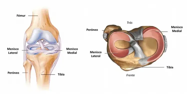
Caption 1: Diagram of both menisci
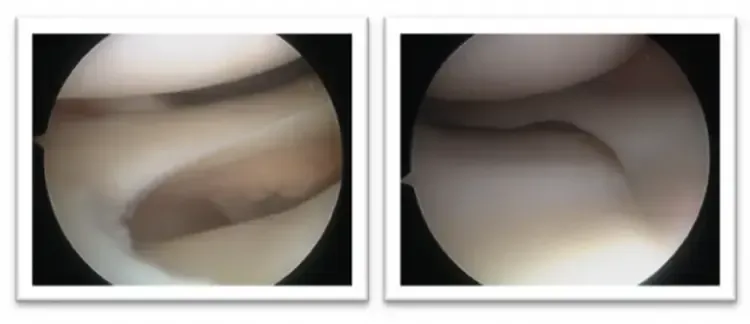
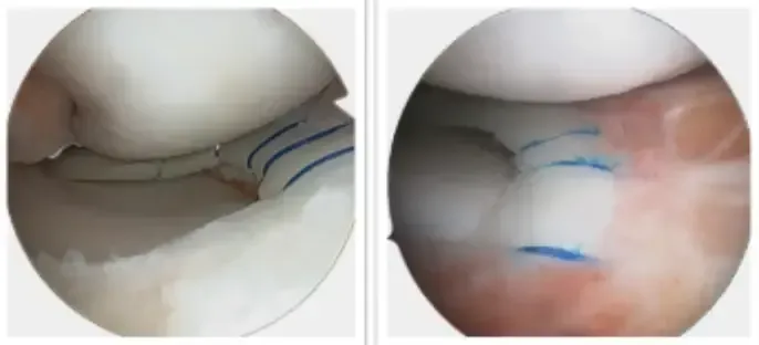
Caption 2: Intra-operative photographs of the medial and lateral meniscus showing normal features
Causes
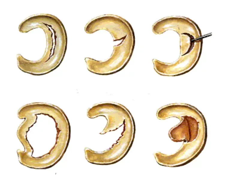
Meniscal tears are among the most common injuries in adult knees. A meniscal tear, also referred to as a "fracture" or simply a meniscal injury, represents a break in the continuity of the meniscus. These tears can occur in various locations or orientations.
While sports-related trauma accounts for the majority of these injuries, they can happen during a variety of activities.
Often, there is an element of knee rotation involved, but up to a third of cases may not involve any notable trauma. This is particularly true in older individuals, where the meniscus's mechanical properties deteriorate over time, increasing susceptibility to injury. In such cases, merely kneeling or squatting might cause a tear.
Symptoms
The most frequent symptom is pain, particularly during rotational movements. This pain typically manifests on the side (either inside or outside) or the back of the knee.
outine activities such as turning over in bed, getting in or out of a car, rotating the leg with the foot planted, crossing the legs, kneeling, or squating can all trigger pain or discomfort. Other symptoms might include swelling, a sensation of instability, or even transient or persistent locking (notably in the case of bucket-handle tears).
Diagnosis
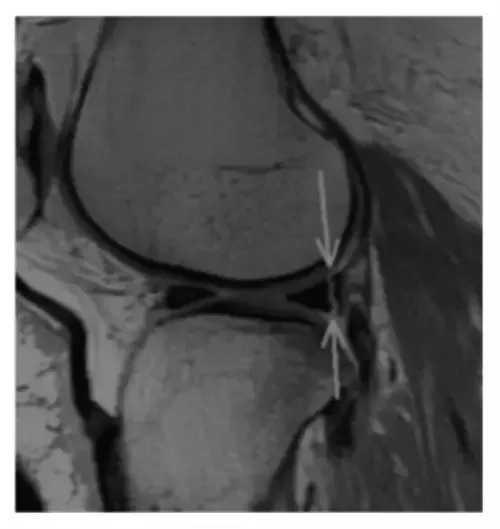
Diagnosis relies on the patient's clinical history, a physical examination, and the results of diagnostic tests. The doctor will check for pain along the femorotibial line and during specific maneuvers. The condition of the knee ligaments will also be assessed, as a meniscal tear often coincides with ligament damage. Magnetic resonance imaging (MRI) is the preferred method for confirming a meniscal tear and provides detailed information about the state of the ligaments and articular cartilage.
While menisci are not visible on X-rays, these images can help rule out associated injuries such as osteoarthritis, fractures, or deviations in knee alignment, which are crucial for determining the most appropriate treatment.
Treatment Options
Not all meniscal tears require surgical intervention. The decision to operate depends on the symptoms presented. Occasionally, a meniscal tear discovered during imaging tests may not be the source of pain. At other times, the tear is only part of the issue (e.g., a degenerative tear in knees with significant osteoarthritis) and should not be addressed in isolation.
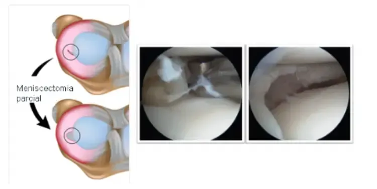
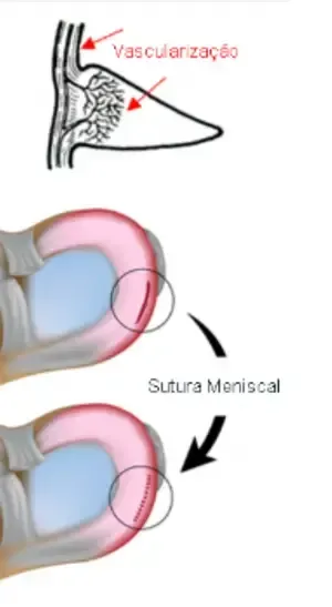
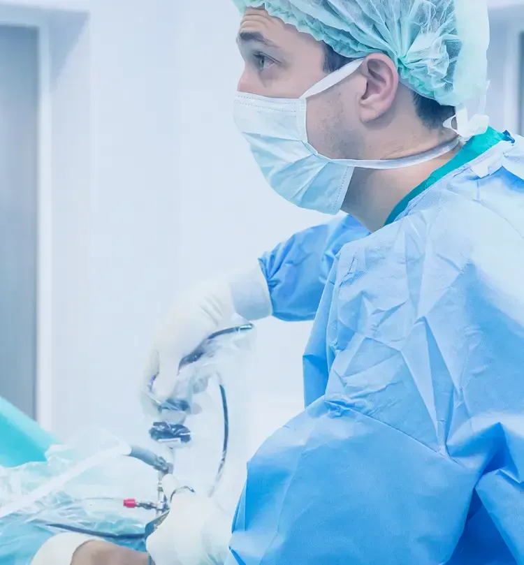
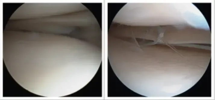
Most isolated meniscal injuries can be effectively managed through simple knee arthroscopy. Based on the tear's characteristics, it can be addressed in one of two ways: partial meniscectomy (removal of the damaged part of the meniscus) or meniscal suturing (repairing the meniscus with stitches to encourage healing).

Only a small fraction of meniscal tears are amenable to suturing, as only the periphery of the meniscus retains blood supply and thus the capacity to heal. In certain cases, treating a meniscal tear may need to be combined with other surgical corrections, such as anterior cruciate ligament reconstruction or osteotomy to correct misalignment of the lower limb.


Anterior Cruciate Ligament Tear
Is one of the key ligaments in the knee that is crucial for maintaining knee stability
Click to find out more information
Copyright © 2026 Ricardo Sousa Ortopedia All rights reserved
Copyright © 2026 Ricardo Sousa Ortopedia
All rights reserved
Powered by Pointfull.


